Dysmorphic uterus (T-shaped uterus) is a congenital uterine anomaly that is common among Thai people with infertility. I once collected research data on every infertility patient who started treatment, using 3D transvaginal ultrasound (3D-TVUS). The prevalence of dysmorphic uterus was as high as 27 percent in this group of infertility patients. I also found that congenital uterine anomalies are an important cause of infertility, and the one most often misdiagnosed (either the anomaly is not detected, or it is detected but diagnosed as another condition), even though treatment is not difficult and, after treatment, nearly 50 percent of patients conceive naturally within the first year.
Normal Uterus
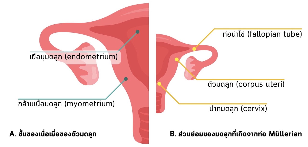
To judge whether a uterus is normal or abnormal, we first need to agree on the angle we look from. A uterus generally looks like two flat sheets of tissue pressed together front to back (this part is called the uterine muscle, or myometrium). The uterine cavity, lined by the endometrium, sits between these two sheets, as shown in Figure 1A.
In addition, in a normal woman of reproductive age, the uterus is about 7 cm long. The body of the uterus is about 4-5 cm long and the cervix about 2-3 cm long, giving a uterine body : cervix ratio of 2:1 (Figure 1B).
Figure 1 A. The tissue layers of the uterus, made up of two main parts: the uterine lining (endometrium) and the uterine muscle (myometrium). B. The parts of the uterus that develop from the Müllerian ducts: the cervix, the body of the uterus (corpus uteri) and the fallopian tubes.
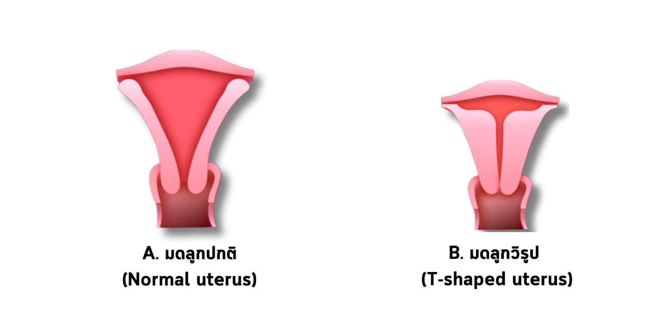
The last feature is the shape of the uterine cavity when viewed front to back (midcoronal view). It looks like an inverted triangle (Figure 2A.), and the uterine muscle surrounding the cavity on three sides is about equally thick.
What is a dysmorphic uterus?
A dysmorphic uterus, or T-shaped uterus, is a congenital uterine anomaly caused by abnormal development of the uterine muscle. A ring-like layer of muscle is too thick, squeezing the uterine cavity until it narrows into the shape of the English letter T (Figure 2B.).
Figure 2 A. A normal uterus. Note that the uterine cavity (brown area) looks like an inverted triangle.
B. A dysmorphic uterus. Note that the uterine cavity is narrower than in a normal uterus and looks like the English letter T. This is because the uterine muscle on the side walls is abnormally thick, squeezing the cavity so it is narrow and small.
A dysmorphic uterus is associated with reduced blood supply to the endometrium, abnormal blood supply to the uterus, abnormal uterine contractions, and abnormal endometrial receptivity, embryo implantation, and expansion of the uterus during pregnancy.
What are the symptoms of a dysmorphic uterus, and which signs suggest higher risk?
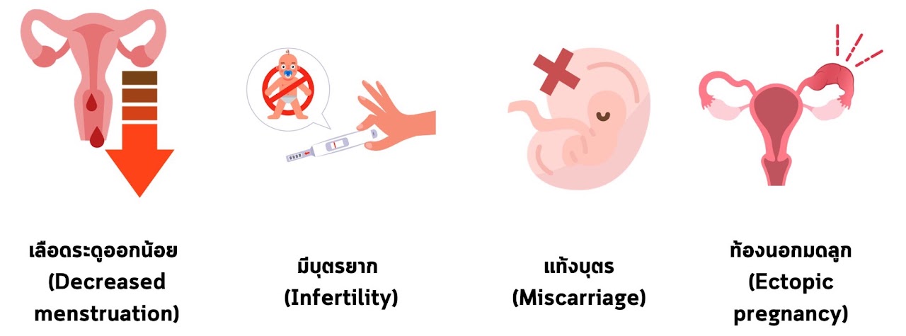
A patient with a dysmorphic uterus who doesn't want children may never be examined and diagnosed, because it usually doesn't cause other gynecologic problems that would bring her in for a check-up. But when it comes to embryo implantation and pregnancy, a dysmorphic uterus lowers the chance of implantation and makes it harder for the uterus to stretch and expand to support the growing baby. So if you have any of the following, you should see a specialist for an assessment, as shown in Figure 3.
Check your own risk for dysmorphic uterus here
Figure 3 Common symptoms or symptom patterns in patients with a dysmorphic uterus (dysmorphic T-shaped uterus)
#LightPeriods Your periods are regular but the flow is light (on your heaviest day you use no more than 3 pads a day, and they aren't even full).
#FirstTrimesterMiscarriage This includes a miscarriage after the baby was seen but had no heartbeat, a blighted ovum, an ectopic pregnancy, or even a blood test that showed you were pregnant (the hCG hormone rose for a while and then fell on its own).
#Hematocrit40PercentOrHigher (Hct). A woman of reproductive age who has a period every month and bleeds a normal amount will have an Hct of about 35-39%. If your blood is more concentrated, at 40% or higher, pay close attention.
#SeeingAFertilityDoctorButStillNotPregnant Whether it's IVF or chromosome testing, it still doesn't work. Here's how I'd put it: normally, 80 percent of women of reproductive age have uterine fibroids, and 50 percent of women with infertility have endometriosis. These causes are easy to find with a regular ultrasound. If your uterus looks very, very normal, that should also make you wonder: normal from which angle? Also, the basic equation for having a baby is a normal embryo + a normal uterus. If you've had normal embryos transferred for a while and still aren't pregnant, I think it's worth looking at the uterus too.
Diagnosing a dysmorphic uterus
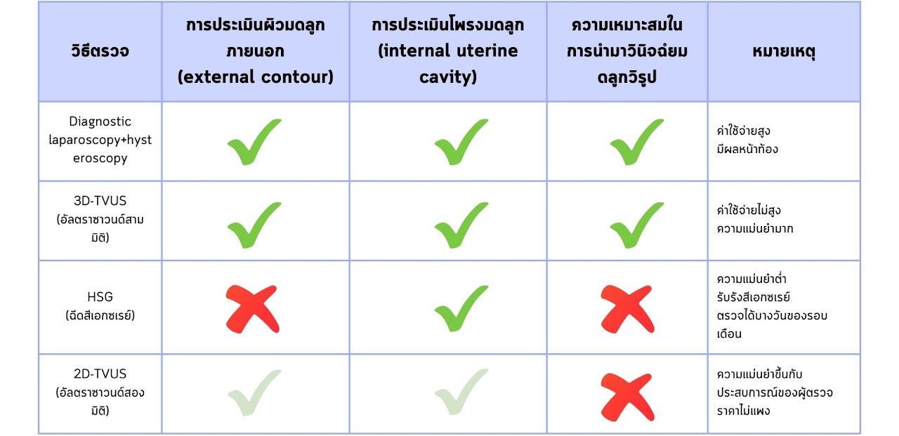
There are several tests to diagnose a dysmorphic uterus. Each has a different sensitivity, and some depend on the examiner's experience, as shown in Table 1.
Table 1 How suitable each test is for diagnosing a dysmorphic uterus (dysmorphic T-shaped uterus)
The diagnostic tool must be able to show the following accurately:
A front-to-back section of the uterus (mid-coronal view)
The external contour of the uterus: it must show whether the outer surface of the uterus is smooth or has an indentation
The shape of the uterine cavity (internal uterine cavity)
Diagnostic hysteroscopy
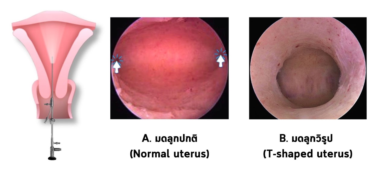
A camera is passed into the uterine cavity through the vagina and cervix, so there is no surgical incision. Today, advances in medical imaging technology mean diagnostic hysteroscopes are only 3-4 mm in diameter. In addition, the vaginoscopy technique (inserting the scope through the vagina) means no speculum or tenaculum (an instrument that holds the cervix) is needed. This makes the procedure more comfortable for patients because there is less pain. A dysmorphic uterus seen through a hysteroscope looks as shown in Figure 4.
Figure 4 A wide-angle (panoramic view) of the uterine cavity through a hysteroscope
A. Normal uterus: the cavity is wide, the lining is pink, and both tubal openings are visible (⬆︎ arrows show the tubal openings).
B. Dysmorphic uterus: the cavity is narrow and deep, like a tunnel, the lining is pale, and the tubal openings cannot be seen.
3D transvaginal ultrasound (3D-TVUS)
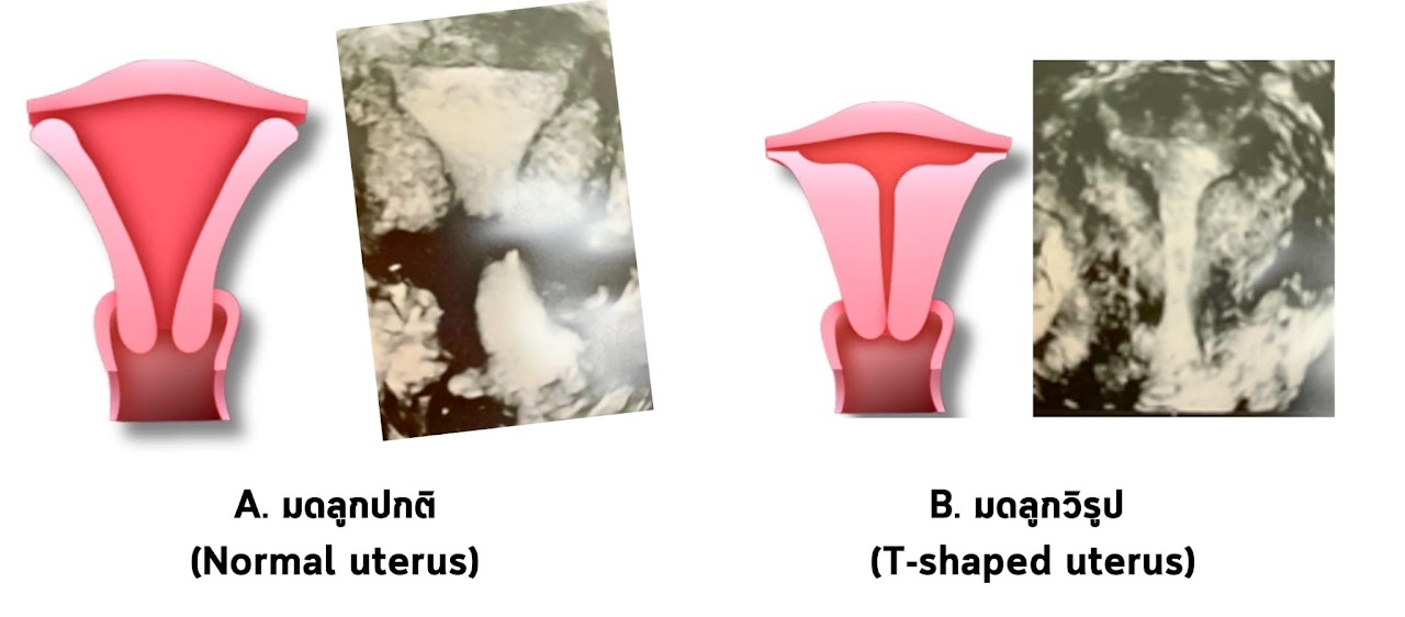
This can clearly adjust the image of the uterus to a front-to-back view (mid-coronal view), so both the external contour and the internal cavity can be assessed with high accuracy. It is inexpensive, and the results are available right away. It does not rely much on the examiner's experience, but it does require extra training in ultrasound skills. A dysmorphic uterus on 3D transvaginal ultrasound looks as shown in Figure 5.
Figure 5 A dysmorphic uterus on 3D transvaginal ultrasound (3D-TVUS)
A. Normal uterus: one uterus with one cavity, and the cavity is triangular.
B. Dysmorphic uterus: one uterus with one cavity. The uterine muscle on the side walls of the cavity is thick, so the cavity is shaped like the letter T.
2D ultrasound (2D-TVUS)
This only shows the uterus from the side (sagittal view), so it is not well suited for diagnosis. The examiner needs advanced ultrasound skills and has to rely on imagination, and the angles in the coronal view cannot be measured, so accuracy is poor.
X-ray dye test (HSG)
This is the same test used to check the fallopian tubes. It can show the internal cavity but cannot show the external contour, so its accuracy is low.
Treating a dysmorphic uterus (Hysteroscopic metroplasty)
Hysteroscopic surgery to restore a normal uterine cavity (Hysteroscopic metroplasty)
There is still no generally accepted standard treatment for dysmorphic uterus, and there are no randomized controlled trials (RCTs) of treatment outcomes. However, experts around the world agree that people with a dysmorphic uterus who want to have children should have surgery through a hysteroscope to restore a normal uterine cavity (hysteroscopic metroplasty). All studies point in the same direction: after surgery, patients have better pregnancy outcomes and fewer miscarriages.
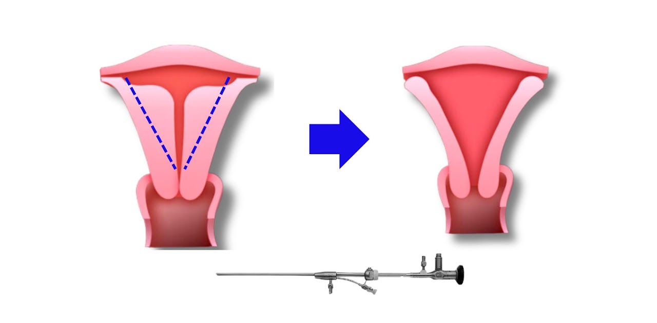
Figure 6 Treating a dysmorphic uterus (dysmorphic T-shaped uterus) with hysteroscopic surgical correction (hysteroscopic metroplasty)
Hysteroscopic metroplasty can be done early in the menstrual cycle, before ovulation (early follicular phase), because the uterine lining is still thin and the uterus does not contract against the instruments. The principle is shown in Figure 6. The left and right uterine muscle is incised in a straight line, and the incision is gradually deepened along that line until the tubal opening (tubal ostium) can be seen when looking up from the isthmus of the uterus. No part of the uterus is removed, unless other abnormalities are found in the cavity at the same time, such as endometrial polyps or submucous fibroids.
Watch a video of hysteroscopic metroplasty here
Care after surgery
After surgery, patients can usually return to almost normal daily life, because the scope is passed through the cervix into the uterine cavity and there is no incision on the abdomen. There may be a little vaginal bleeding, especially in the first week.
We recommend avoiding sex for 1 month after surgery, or until your next period comes, because it could cause infection and inflammation of the uterine cavity.
Adhesions inside the uterine cavity after surgery can be prevented by measures such as placing the incisions on the left and right sides, where scarring is very unlikely; placing an anti-adhesion gel in the cavity right after surgery; taking high-dose estrogen by mouth to stimulate the lining to grow over the surgical area; and preventing infection after surgery with antibiotics.
Follow-up after treatment
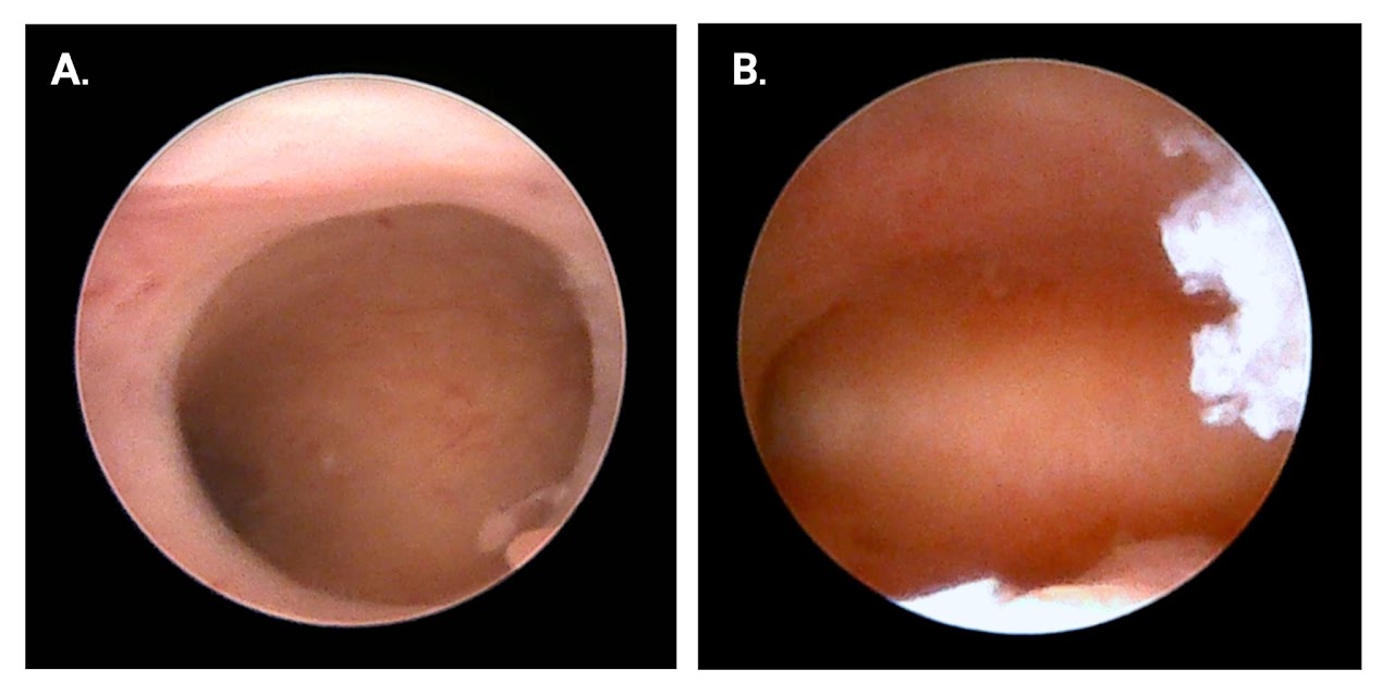
After surgery, the shape of the uterine cavity should be checked again with diagnostic hysteroscopy or 3D transvaginal ultrasound (3D-TVUS) to see whether it has returned to normal. Diagnostic hysteroscopy with the vaginoscopy technique can be done as an outpatient procedure and doesn't take long. Patients feel little or no pain (Figure 7).
Figure 7 The uterine cavity on diagnostic hysteroscopy in the same patient with a dysmorphic uterus. A. The cavity before surgery: note the pale lining, the narrow, deep cavity and the tubal openings that cannot be seen. B. The cavity 5 weeks after hysteroscopic metroplasty, showing a normally wide cavity, a lining with normal blood supply, and both tubal openings visible.
After the post-surgery check, you can try to conceive naturally or start ovarian stimulation for IVF right away. If you plan a frozen embryo transfer, we recommend waiting at least 2 menstrual cycles before starting to prepare the uterine lining for the frozen embryo transfer.
Advice before and after hysteroscopy
Outcomes of dysmorphic uterus treatment after surgery
Hysteroscopic correction of a dysmorphic uterus with the Digital hysteroscopy clinic technique makes hysteroscopic surgery safer, more precise and more convenient. It restores a normal uterine cavity, ready to receive an embryo. Over time, nearly a hundred patients have had this surgery here, so I gathered the pregnancy outcomes of patients with a dysmorphic uterus after surgery. Let's take a look.
We found that people whose dysmorphic uterus had been corrected had a pregnancy rate of 72.5% within the first year after surgery.
After surgery, 66.7% of patients conceived naturally
33.3% of the patients who conceived needed assisted reproductive technology (IVF, ICSI)
We found that whether patients conceived naturally or through IVF, it took a little over 5 months.
Summary
Dysmorphic uterus (dysmorphic T-shaped uterus) is a congenital uterine anomaly that is common in people with infertility, and it is a cause of infertility and miscarriage.
Patients often have light periods (no more than 3 pads on the heaviest day, and periods lasting fewer than 4 days), repeated miscarriages, or no pregnancy after IVF and transfer of chromosomally normal embryos.
Diagnosis uses 3D ultrasound (3D-TVUS) or hysteroscopy.
Treatment is hysteroscopic surgery to reshape the uterine cavity (hysteroscopic metroplasty).
Surgery clearly increases the chance of pregnancy and lowers the chance of miscarriage.
References
Patsama Vichinsartvichai. Chapter 11: Dysmorphic uterus. In: Patsama Vichinsartvichai. Congenital malformation of uterus [in Thai]. 1st edition, B.E. 2565 (2022).
Coelho Neto MA, Ludwin A, Petraglia F, Martins WP. Definition, prevalence, clinical relevance and treatment of t-shaped uterus: Systematic review. https://doi.org/10.1002/uog.23108. Ultrasound in Obstetrics & Gynecology. 2021/03/01 2021;57(3):366-377.
Leave a comment
* This article is general health education. Diagnosis and treatment should be guided by your doctor. Results vary with age and individual medical factors.
Dr. Patsama Vichinsartvichai, MD, MClinEmbryol, EFOG-EBCOG, EFRM-ESHRE/EBCOG, FACOG is a board-certified reproductive endocrinology & infertility specialist, former head of the Vajira IVF unit at Navamindradhiraj University, and founder of LIFE by Dr. Pat in Bangkok. His published research focuses on the dysmorphic (T-shaped) uterus and uterine-factor infertility.
 LIFE by Dr. Pat