Pre-implantation genetic testing for aneuploidy (PGT-A)
Diploma in Gynecologic Endoscopic Surgery, MClinEmbryol,
EFOG-EBCOG., EFRM-ESHRE/EBCOG.
Introduction
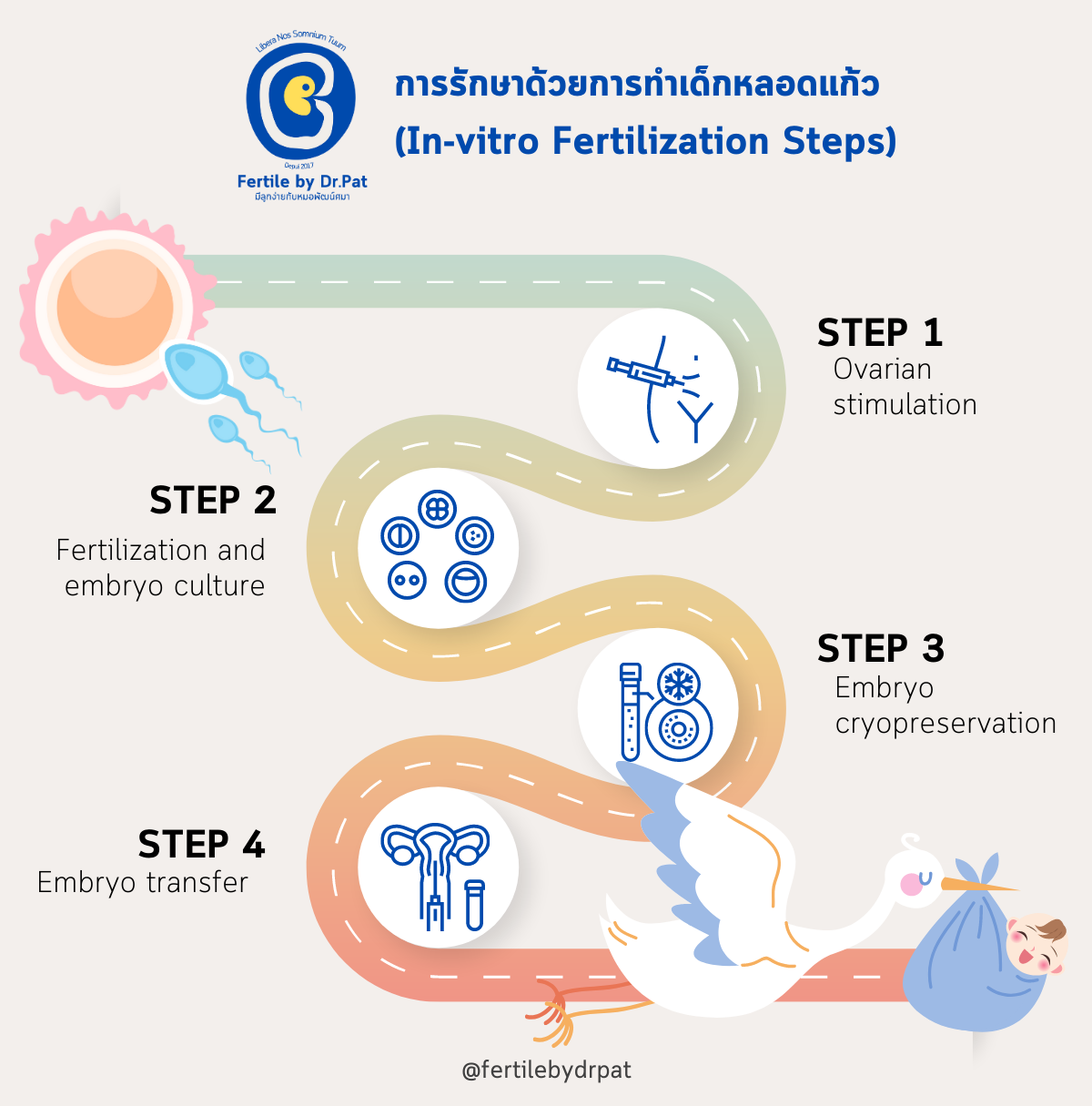
Many couples with infertility need IVF treatment (IVF/ICSI cycles), a complex treatment with many steps, as summarized in Figure 1. Choosing which embryo to transfer (embryo selection for transfer) is one of the steps that plays an important role in pregnancy outcomes, and there is still no clear conclusion on which embryo selection method or process is better than the others.
Figure 1. The steps in IVF treatment (Steps in IVF/ICSI treatment)
How to Choose the Embryo with the Best Chance of Implanting
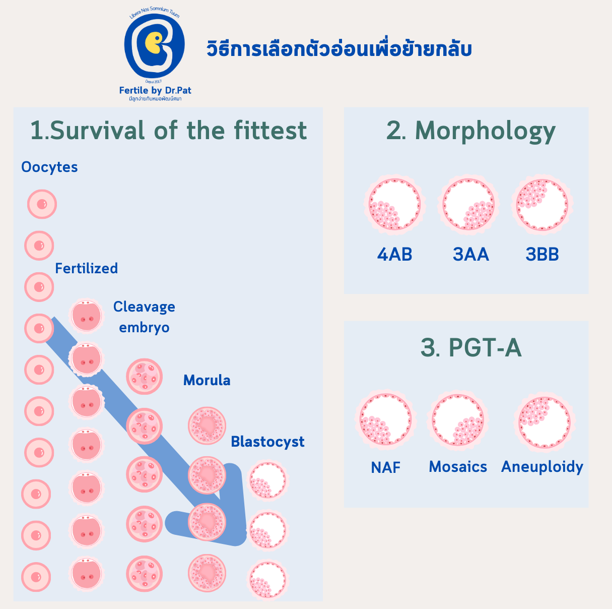
Several strategies are used to select the embryo most suitable for transfer, as shown in Figure 2.
Figure 2. Strategies used in today's embryology laboratories to select embryos for transfer: 1) Survival of the fittest 2) Morphology, the shape and appearance seen under the microscope 3. Embryo chromosome testing (PGT-A)
Survival of the fittest
#TheWeakFallAway. This is the principle nature uses to select automatically, without favoritism. In fact, it is also how a natural cycle decides which egg will be ovulated. (A woman has follicles that are ready to develop into the follicle that will go on to ovulate, which we call antral follicles. Normally 10-20 antral follicles start to grow, but only 1 develops all the way to ovulation. The other antral follicles degenerate and die off on their own through a process called atresia.)
As an embryo develops from a fertilized egg to the blastocyst stage (day 5-6 after ovulation), some do not reach the finish line along the way. For example, only 60-80% of inseminated eggs are actually fertilized, and only 60-70% of fertilized eggs develop into blastocysts. Roughly speaking, only 30-40% of the mature eggs collected develop into usable blastocysts.
Morphology
This means assessing embryo quality under the microscope, using criteria such as the number of pronuclei (day 1), the number of cells in the embryo and how even the cells are in shape, the proportion of fragments (cell debris) in the embryo (day 3), and, at the blastocyst stage, the appearance of the trophectoderm and inner cell mass and how expanded the blastocyst is. However, selecting embryos by morphology works only partly. Only 1 in 3 patients who have an embryo transfer selected by morphology become pregnant, and many of them still miscarry. This is most likely because the embryos have an abnormal number of chromosomes.
I like to compare embryo development to a #BeautyPageant. Each day, some embryos go on to develop and some don't move on, just like contestants being eliminated in each round. You can read more about embryo grading in this article.
PGT-A
We know that with embryos, #YouCantJudgeABookByItsCover. How an embryo looks does not match whether its chromosomes are normal, and chromosome abnormalities cause embryos not to implant or to miscarry. So the last method is chromosome testing, which is what this article is about. A few cells are removed from the embryo for genetic analysis to identify how many chromosomes, and how many sets, those cells contain.
Only embryos with a normal number of chromosomes move on to the next round and are transferred into the uterus.
That sounds very reasonable, doesn't it? It even seems to bring a glimmer of hope.
Why Test Embryo Chromosomes?
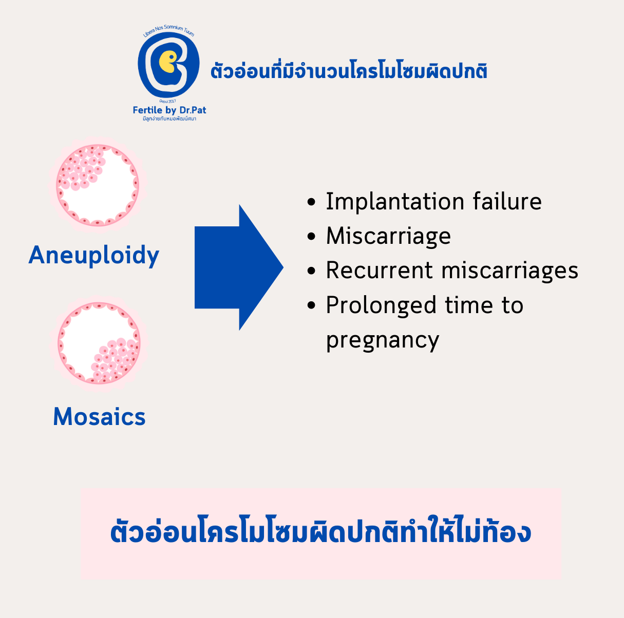
Figure 3. The basic principle behind the research and development of preimplantation chromosome testing of embryos (PGT-A)
We know that most normal-looking embryos that are transferred and do not result in pregnancy have an abnormal number of chromosomes. In addition, chromosomally abnormal embryos increase the chance of miscarriage and lengthen the time from the start of treatment to pregnancy (Figure 3). In other words, #WastedTime.
Does a Chromosomally Normal Embryo Mean the Baby Will Be Normal?
Here is my short answer first...
It doesn't follow
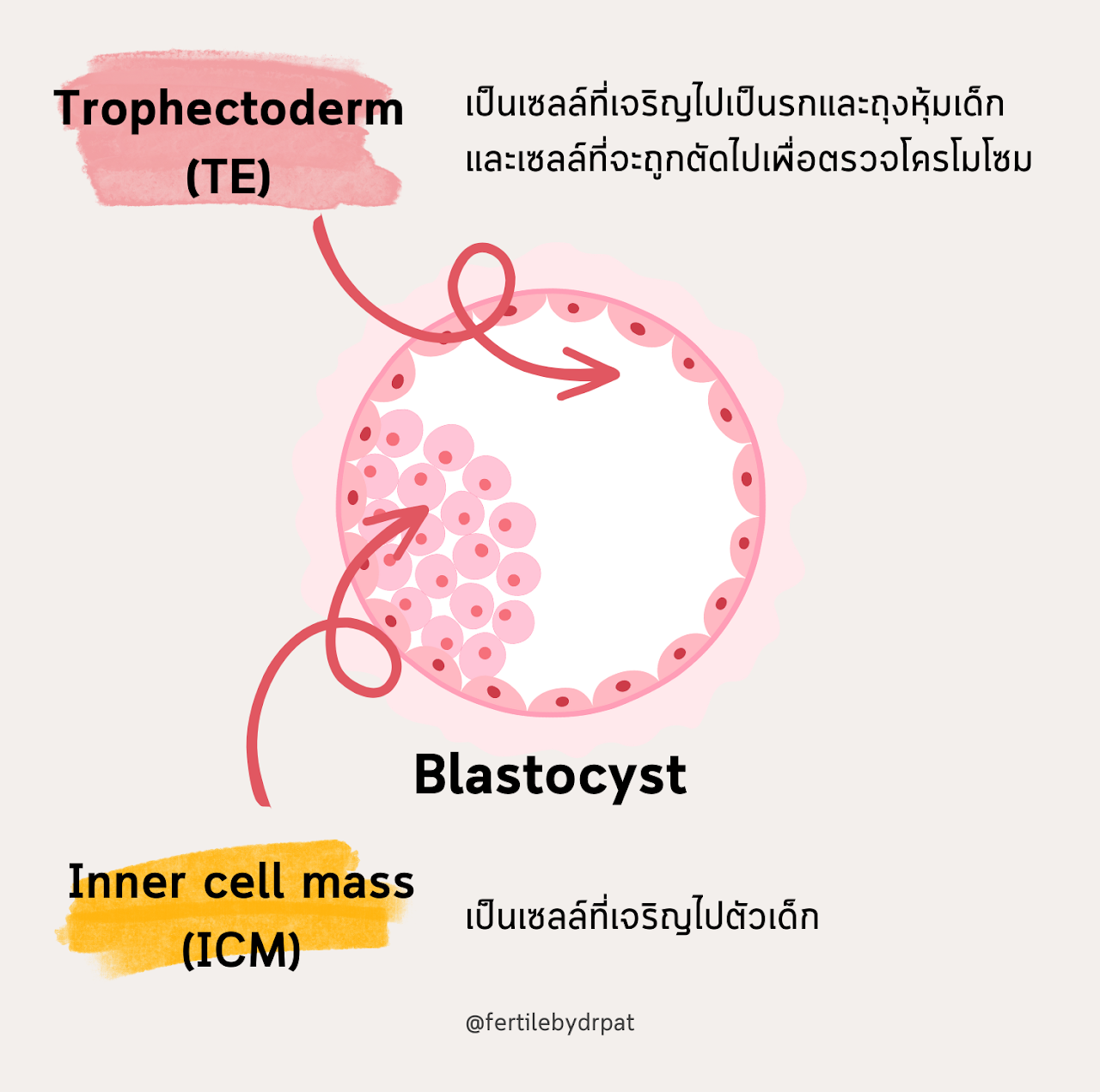
Figure 4. The parts of an embryo at the blastocyst stage (day 5-6 after ovulation)
Today, the embryos we biopsy are usually at the blastocyst stage (day 5-6 after ovulation), which is made up of the cells shown in Figure 4. Five to 10 trophectoderm cells are removed and sent for chromosome testing. These are not the cells that will develop into the baby. And remember that as the embryo develops, its cell number doubles within a single day. So the cells that will become the baby may have chromosomes that differ from those in the trophectoderm, although the chance of this is not high.
So the purpose of PGT-A is to help patients get pregnant sooner, not to prevent chromosome abnormalities in the baby. PGT-A cannot replace prenatal screening for chromosome abnormalities in the baby. A normal chromosome result does not mean the baby born from that embryo will be normal.
"The purpose of PGT-A is to help patients get pregnant sooner
It cannot replace prenatal screening for chromosome abnormalities in the baby
A normal chromosome result does not mean the baby born from that embryo will be normal"
Treatment Outcomes
What do patients expect from PGT-A? A higher chance of pregnancy? A healthier baby? Can these really happen, or is the data misleading us? Let me explain.
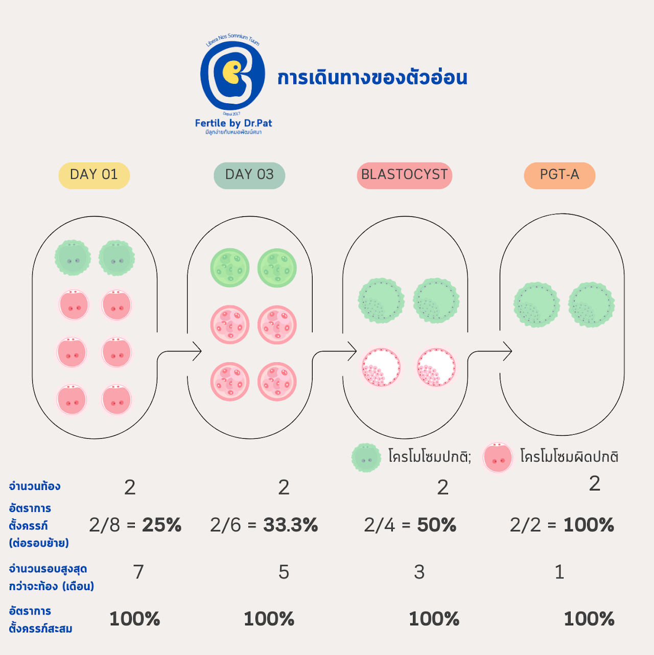
Figure 5. An example of a patient's embryo development from fertilization to day 5-6 after ovulation, with PGT-A, and hypothetical treatment outcomes if embryos were transferred at different stages
Figure 5 shows hypothetical data on embryo development in an IVF cycle, calculated from commonly used competency and benchmarking KPIs.
Humans are the only species in the world in which an embryo transferred into the uterus at any stage can implant and lead to a live birth. Today's medical technology lets us freeze and thaw embryos with very little damage to them. Biopsying cells from a blastocyst-stage embryo also has very little effect on implantation. And culturing embryos outside the body up to the blastocyst stage is considered to allow better embryo selection.
It all sounds good, doesn't it?
But don't forget that culturing embryos outside the body for several days, removing some of their cells, and freezing and thawing them do not make the embryos better quality. Even in the hands of the most skilled medical team, careful handling at each of these steps can, at most, keep the embryo close to the quality it already had. If anything goes wrong, the embryo's quality will get worse. So my conclusion number 1 is:
"Embryo chromosome testing does not make embryos better
(they stay the same, or get worse if the people handling them are not skilled)"
Since embryos that go through these processes do not get better, testing does not increase the number of pregnancies. Put simply, it does not give you more children. We may believe chromosome testing raises the chance of pregnancy because of pregnancy rates per embryo transfer. This is the same reason we believe blastocyst transfer gives higher pregnancy rates. Without testing, embryos with a low chance of implantation are also transferred, so when the data are pooled per cycle, implantation and pregnancy rates per transfer are low.
But judging the data this way isn't fair. If we transfer all of one patient's embryos until none are left, we find that the number of children from treatment is no different with or without chromosome testing. But the time to pregnancy will be shorter if the embryos are tested (all shown in Figure 5). This is called the cumulative pregnancy rate, and it is a more important measure than the pregnancy rate per embryo transfer, which reflects only that one treatment cycle.
"PGT-A does not increase pregnancy rates
but it helps you get pregnant sooner"
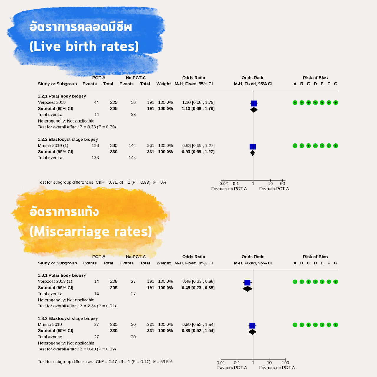
Now let's see whether the research matches my assessment. A Cochrane review by Cornelisse and colleagues (1), which pooled data from several RCTs, found that with current PGT-A technology, live birth rates did not differ between testing and not testing, and neither did miscarriage rates, as shown in Figure 6.
Figure 6. Live birth rates and miscarriage rates compared between PGT-A and no PGT-A
Reference: Adapted from Cornelisse S, et al. Preimplantation genetic testing for aneuploidies (abnormal number of chromosomes) in in vitro fertilisation. Cochrane Database Syst Rev 2020.
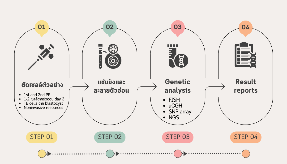
Laboratory Steps in Embryo Chromosome Testing (PGT-A)
Figure 7. The laboratory steps in PGT-A
Summary
Figure 8. Summary of where PGT-A stands based on the data available in 2023 (B.E. 2566)
References
Cornelisse S, et al. Preimplantation genetic testing for aneuploidies (abnormal number of chromosomes) in in vitro fertilisation. Cochrane Database Syst Rev 2020.
ESHRE Working Group on Recurrent Implantation Failure and others, ESHRE good practice recommendations on recurrent implantation failure, Human Reproduction Open 2023. https://doi.org/10.1093/hropen/hoad023
The ESHRE Guideline Group on RPL and others, ESHRE guideline: recurrent pregnancy loss: an update in 2022, Human Reproduction Open, Volume 2023, Issue 1, 2023, hoad002, https://doi.org/10.1093/hropen/hoad002
The Unexplained Infertility guideline group, Romualdi D, Ata B, Bhattacharya S, Bosch E, Costello M, Gersak K, Homburg R, Le Clef N, Mincheva M et al. Evidence-based guideline: Unexplained Infertility 2023. ESHRE, https://www.eshre.eu/guideline/UI
Angell RR, Aitken RJ, Van Look PF, Lumsden MA, Templeton AA. Chromosome abnormalities in human embryos after in vitro fertilization. Nature 1983;303(5915):336-8. [DOI: 10.1038/303336a0]
* This article is general health education. Diagnosis and treatment should be guided by your doctor. Results vary with age and individual medical factors.
Dr. Patsama Vichinsartvichai, MD, MClinEmbryol, EFOG-EBCOG, EFRM-ESHRE/EBCOG, FACOG is a board-certified reproductive endocrinology & infertility specialist, former head of the Vajira IVF unit at Navamindradhiraj University, and founder of LIFE by Dr. Pat in Bangkok. His published research focuses on the dysmorphic (T-shaped) uterus and uterine-factor infertility.
 LIFE by Dr. Pat